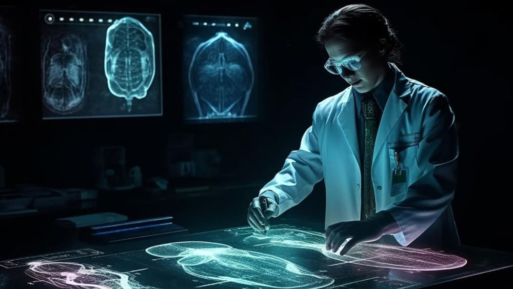
AI-powered radiology is ushering in a new era where diagnostic speed and accuracy no longer compete but complement each other.
AI-Powered Radiology: Speeding Up and Improving Diagnostic Accuracy
Written by Sumit Kaushik

Radiology, which is one of the pillars of diagnostic medicine, has in the past relied on the human factor of competent radiologists to interpret medical images. However, with imaging study boom on all sides and disease presentation complexity worldwide increasing, the traditional model is straining at the seams.
Artificial Intelligence (AI), in the form of machine learning and deep learning, is increasingly playing the lead role as the game-changer—a paradigm-shifting element that is bringing historic speed, accuracy, and efficiency to radiologic diagnoses.
This marriage of medicine and technology is revolutionizing the practice of diagnosis, detection, and treatment of disease in the healthcare industry, which, in turn, influences patient outcomes.
The Future Issue in Radiology: Data Overload and Diagnostic Complexity
New radiology produces stunning volumes of data every day. Imaging centers, hospitals, and diagnostic laboratories produce millions of images—X-rays and CT scans, MRIs, and PET scans. A single scan can take hundreds to thousands of images that have to be carefully read.
Radiologists have to spot minute variations in these images—minute nodules, microfractures, minuscule hemorrhages, or slight changes in the tissue. Delayed or missed diagnoses mean serious health consequences. Also, the global shortage of radiologists worsens the condition, leaving additional volumes and fatigue factors vulnerable.
AI here is not only a luxury but a requirement to help deal with the amount and diversity of today's imaging diagnostics.
The Radiology Artificial Intelligence Technology: Machine Learning and Deep Learning
All but a few artificial intelligence algorithms employed in radiology are machine learning (ML) and more sophisticated deep learning (DL), a subset of ML that learns from patterns in the neural networks of the human brain. Artificial intelligence is trained on vast annotated collections of medical images and identifies patterns and features akin to various pathologies.
- Convolutional Neural Networks (CNNs): Most appropriate for image recognition applications. CNNs can handle pixel data and can be trained to recognize features such as texture, edge, and shape—critical to recognize anomalies in scans.
- Natural Language Processing (NLP): Utilized for composing or assisting in composing clinical reports by converting radiologist remarks and mapping image findings.
- Reinforcement Learning: Some of the more advanced models have AI trained in feedback loops to enhance diagnostic accuracy with each successive block of time.
The continuous training and refinement over multiple datasets enable AI to build its sensitivity (detection of genuine positives) and specificity (avoidance of false positives), which are critical for clinical acceptability.
Key Benefits of AI in Radiology
- Optimized Diagnostic Workflow: AI is capable of reviewing scans in real-time as they are being created, priority case by priority case. Therefore, such critical detections like intracranial hemorrhage, pulmonary embolism, or bowel obstruction are identified instantly, and immediate intervention can be offered. AI is a fast screen that allows radiologists to manage tasks efficiently and optimize patient flow.
- Enhanced Diagnostic Accuracy: AI excels at recognizing subtle and early-stage disease features that may escape human detection. For example, early lung nodules or breast microcalcifications may be too faint or obscured for even expert eyes but can be identified by AI’s pattern recognition capabilities.
- Reduction of Human Error: Fatigue, inattention, or going outside experience can lead to errors in missed diagnoses or misinterpretation. AI provides a consistent, uncompromising second opinion minimizing diagnostic error and inter-reader variability and overall quality of care optimization.
- Quantification and Monitoring Using Automation: Radiology often is accurate measurement—tumor size, organ volume, vessel diameters. AI can simply automate them with high reproducibility, enabling uniform monitoring of disease progression or response to therapy.
- Enabling Personalized Medicine: AI integrates imaging information with demographics, patient history, genetic information, and laboratory tests to create a comprehensive patient image. Multi-dimensional analysis enables precision diagnosis and customized treatment plan.
Clinical Application of AI-Based Radiology
Oncology
Lung, breast, and prostate cancers are pre-clinically identified by AI algorithms to find suspicious lesions with extremely high sensitivity.
Tumor segmentation and volumetry automatically assist oncologists with treatment planning and response evaluation.
Neurology
Stroke is a matter of time. Ischemic stroke and hemorrhage on CT or MRI are rapidly diagnosed using AI tools such that thrombolytic therapy or surgery can be performed with the earliest.
Cardiology
Cardiac imaging is scanned by artificial intelligence for coronary artery disease, plaque composition, and myocardial function evaluation.
The detection for arrhythmogenic substrates is improved and risk stratification is simplified.
Orthopedics and Trauma
AI-assisted hairline fracture, joint disease, and degenerative illness diagnosis diminishes missed injury in the emergency room.
Infectious Disease and Pulmonology
AI was used in the COVID-19 pandemic in the detection of widespread lung abnormality on chest CT and tracking of disease progression.
Limitations for AI Adoption in Radiology
- Data Privacy and Security: Health data is personal and tightly regulated. Patient information needs to arrive in batches to train and test AI systems, which compounds fears of privacy infringement. Approaches like federated learning allow AI systems to learn from heterogeneous data without violating patient confidentiality.
- Bias and Generalizability: AI trained on restricted or homogeneous data can be poor performers in heterogeneous populations, exacerbating health inequities. Ongoing work at inclusive, multi-center data collections will be necessary to developing equitable AI models.
Radiologist Trust and Workflow Integration: Adoption depends on radiologists being able to take AI results and integrate them into work flows without stalling them. AI will have to be accepted as an assistant, not a replacement. Training must develop competence and confidence.
Regulatory Approval and Liability: AI diagnosis software are subjected to extensive testing and approval by regulatory authorities (FDA, EMA). Being held responsible if an AI fails to diagnose or misdiagnoses still remains an ethical as well as a legal challenge.
The Future: Cooperative Intelligence and Beyond
AI is not replace radiologists. The future is cooperative intelligence, when man's intellectual power and machine's are harnessed by means of synergy.
Explainable AI
Explainable AI design enhances clinician trust and transparency. Radiologists are better informed when making decisions about AI recommendations with explanations.
Multimodal Diagnostics
Next-generation AI will combine imaging with genomics, pathology, lab tests, and clinical data, producing more detailed diagnostic information than one image interpretation.
Real-Time AI Integration
AI embedded in image devices will provide real-time image acquisition feedback to improve quality and reduce repeat scans.
Personalized Treatment Pathways
Predictive AI analytics will guide individualized treatment regimens, predicting the outcome on the basis of imaging phenotypes and clinical factors.